Mèlanie Reijnaers
Cancer, being the second leading cause of death worldwide, is a major public health problem [1]. Every day, thousands of researchers are trying to make a difference; to help the patients fight the devastating effects the disease brings. This research is not only focussed on trying to create new treatments, but also on figuring out the behaviour of the disease. Recently, in august 2020, Ubellacker et al. revealed the advantage that melanoma cells receive when they migrate through the lymphatic system instead of the blood system [2]. This phenomenon is believed to have a pivotal role in the prognosis of cancer. For this reason, the intriguing mechanism behind the advantage will be discussed in this brief message, highlighting the importance of further research on this topic.
The migration of cancer cells from one tissue to another, leading to the formation of secondary tumours, is known as metastasis. Metastasis can occur through multiple routes of which the primary ways are transcoelomic spread (via the abdominal cavity), hematogenous spread (via the blood), lymphatic spread, and a combination of the latter two. Even though metastasis is common in cancer patients, the process is known to be inefficient as not many cells survive in the blood [3, 4]. The main reason for this inefficiency is the presence of oxidative stress in this vital fluid [5]. As you might have already learnt during your studies, oxidative stress can induce cell death in multiple ways. Ubellacker et al. observed in in vivo assays that the melanoma cells are killed in the blood via a process called ferroptosis, which is induced by oxidative stress [2].
Ferroptosis is an iron-dependent form of cell death and causes lipid peroxidation. Polyunsaturated fatty acids (PUFAs) are in particular sensitive to the oxidative mechanism [6]. The rule goes as follows: the more unsaturated a phospholipid is, the more prone it is to undergo ferroptosis [7]. The oxidation of these PUFAs is the main reason why cells that transit through the lymphatic system have an advantage in spreading compared to cells that travel via the blood. To understand why this is the case, it is essential to know that the cell membrane usually contains many PUFAs. However, what is the reason for the advantage for cells that have been in the lymphatic system? Why does this milky-white, lymphocyte rich, fatty fluid lead to a beneficial effect for metastasis?
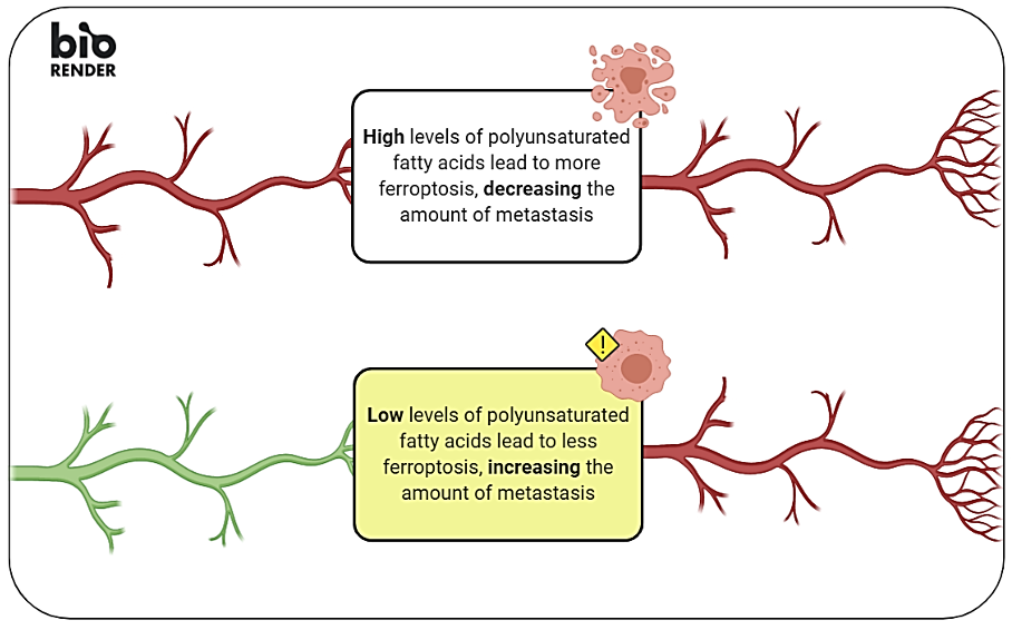
Well, that the lymphatic fluid is rich in triglycerides, alias fatty, is mostly the reason. Ubellacker et al. observed that malignant cells that entered the lymphatic system were significantly more enriched with oleic acid, a monounsaturated fatty acid (8.7 ± 0.9-fold (mean ± standard deviation)) [2]. This oleic acid, as well as other monounsaturated acids, acts as an inhibitor for ferroptosis by replacing and thereby reducing the density of PUFAs in the cell membrane. The reduced density of PUFAs leads to fewer available sites that can undergo ferroptosis. Therefore, when malignant cells enter the blood after they have been in the lymph nodes/vessels, the process of ferroptosis is avoided [2]. Subsequently, these cells have a higher chance to survive in the bloodstream leading to an increased risk of metastasis and a worse prognosis of the disease [Figure 1].
This result was supported by additional results showing that melanoma cells in the presence of ferroptosis-inhibitor molecules led to the same amount of metastasis as the malignant cells that entered the lymphatic system without the inhibitor, suggesting that such cells did not undergo ferroptosis [2]. Moreover, the study of Ubellacker et al. measured a higher concentration of malignant cells in the lymphatic system compared to the blood system, which indicates as well that the fatty environment of the lymph contributes to an anti-cell-death mechanism [2]. These findings, and many more results from the study of Ubellacker and colleagues, provide a deeper understanding of the behaviour of malignant cells and lay the groundwork for future research into the protective environment of the lymph.

References:
[1] Anonymous. Global, regional, and national life expectancy, all-cause mortality, and cause-specific mortality for 249 causes of death, 1980-2015: a systematic analysis for the Global Burden of Disease Study 2015. Lancet (London, England)388, 1459-1544 (2016).
[2] Ubellacker, J.M., et al. Lymph protects metastasizing melanoma cells from ferroptosis. Nature 585, 113-118 (2020).
[3] Mehlen, P. & Puisieux, A. Metastasis: a question of life or death. Nature reviews. Cancer 6, 449-458 (2006).
[4] Vanharanta, S. & Massagué, J. Origins of metastatic traits. Cancer cell24, 410-421 (2013).
[5] Piskounova, E., et al. Oxidative stress inhibits distant metastasis by human melanoma cells. Nature 527, 186-191 (2015).
[6] Dixon, S.J. & Stockwell, B.R. The Hallmarks of Ferroptosis. Annual Review of Cancer Biology 3, 35-54 (2019).
[7] Spitz, D.R., et al. The effect of monosaturated and polyunsaturated fatty acids on oxygen toxicity in cultured cells. Pediatr Res 32, 366-372 (1992).
